
New corneal research suggests a path for studying EyeCell™ Plus and bioelectric stimulation in tissue repair.
A 2026 study in Nature Communications offers a striking view of how the eye can repair itself. After researchers removed stem cells from the surface of mouse corneas while preserving the surrounding tissue environment, mature corneal cells moved into the emptied area and acquired characteristics of limbal stem cells. The regenerated cells supported long-term surface renewal and repaired subsequent injuries. The researchers also found that signals from the local cellular environment could encourage stem cell features in cultured human corneal cells.
The finding points to a new question for regenerative medicine: Can a treatment help create the conditions in which a person’s own cells repair tissue? That question fits Lionheart Health’s interest in bioelectric signals, targeted stimulation, and multimodal protocols. It does not mean that EyeCell™ Plus has been shown to reproduce the study’s results.
The local environment matters
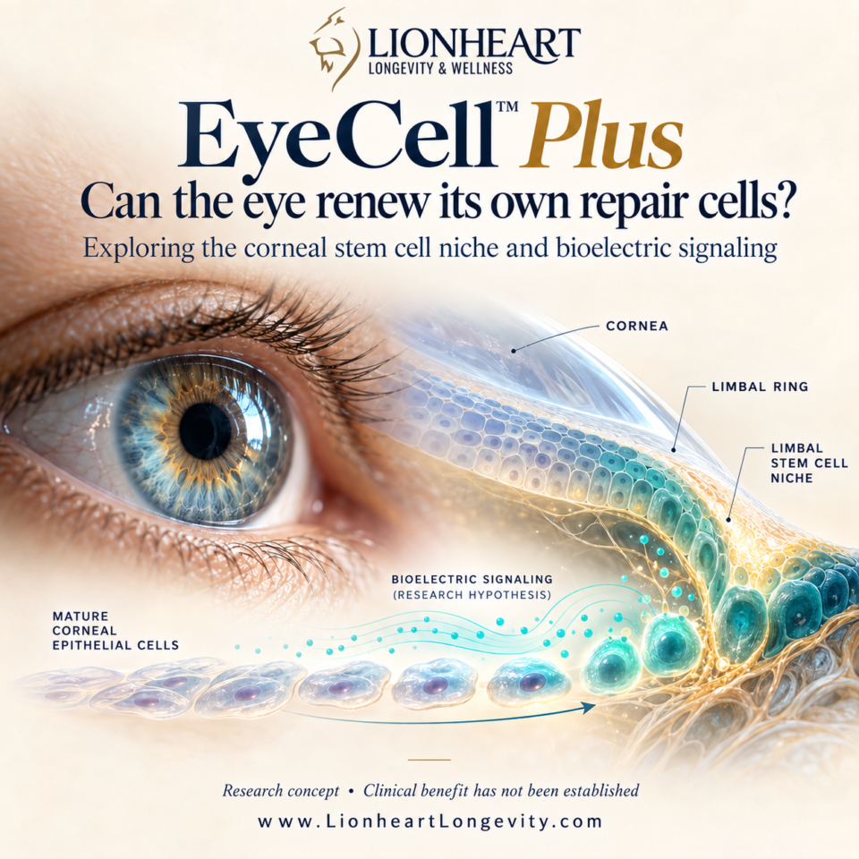
The study did not show that any mature cell can become any kind of stem cell. The response depended on an intact limbal niche: the local environment that supports the cornea’s stem cells. Corneal epithelial cells contributed to regeneration, while cells from the neighboring conjunctiva did not replace the corneal stem cell population. The researchers identified signals from cells in that niche as part of the process.
For Lionheart, this suggests a specific research direction. EyeCell™ Plus could be studied for whether its stimulation changes measurable features of the ocular surface environment, including repair related signaling, inflammation, and epithelial recovery. Bioelectric stimulation might eventually prove useful as one component of a protocol designed to support a healthy niche. That is a hypothesis to test, not an outcome established by this paper. The study did not test EyeCell™, the Lionheart 240 Stimulus™, or electrical stimulation.
From a promising mechanism to a testable program
A useful next step would be to study Lionheart’s proposed approach in stages. Laboratory work with human corneal epithelial cells could examine whether defined stimulation settings affect cell survival, movement, gene expression, and stem cell associated markers. Further research could assess whether any changes are beneficial, durable, and safe for the eye. Clinical studies would then need to measure outcomes that matter to patients, such as ocular surface healing and visual function.
The paper also gives researchers an important boundary. Its strongest evidence for sustained regeneration comes from mouse corneas; the human cell experiments support the relevance of the signaling pathway but do not establish a treatment for patients. Any claim that a Lionheart protocol restores limbal stem cells would require direct evidence from Lionheart’s own studies.
A broader idea for regenerative health
Lionheart’s technology platform is built around the possibility that precisely delivered signals can influence how tissue responds. This corneal study makes that idea worth investigating in a more focused way: preserve or support the right local environment, measure the signals cells receive, and test whether those signals improve repair.
The immediate opportunity is a research program, not a promised clinical result. If Lionheart can show that EyeCell™ Plus safely improves the conditions for ocular surface repair, this work could help guide future protocols. The larger lesson from the study is encouraging: even differentiated cells may retain more capacity to contribute to repair than previously assumed, when the right tissue conditions are present.
Research perspective. EyeCell™ Plus has not been shown in this study to convert mature corneal cells into stem cells or to treat limbal stem cell deficiency.