
Repairing bone requires a coordinated biological response. Cells must reach the injury, multiply, establish a blood supply, build a mineralized scaffold, and remodel that scaffold into strong, functional tissue.
Lionheart Health and Leonhardt Ventures are investigating whether programmed bioelectric signals can influence proteins involved in these processes. Their publicly described intellectual-property portfolio includes issued patents, published applications, and developing research concepts involving bone formation, stem-cell recruitment, blood-vessel support, and tissue remodeling. Published implant-healing applications describe this proposed bioelectric approach. Patent protection, however, does not itself establish clinical effectiveness.
The scientific opportunity is to influence the right biological signals at the right location and stage of healing. Several proteins offer compelling research targets, while others require particular care because their effects can support or inhibit bone formation depending on context.
Tropoelastin: Helping Repair Cells Attach, Multiply, and Move
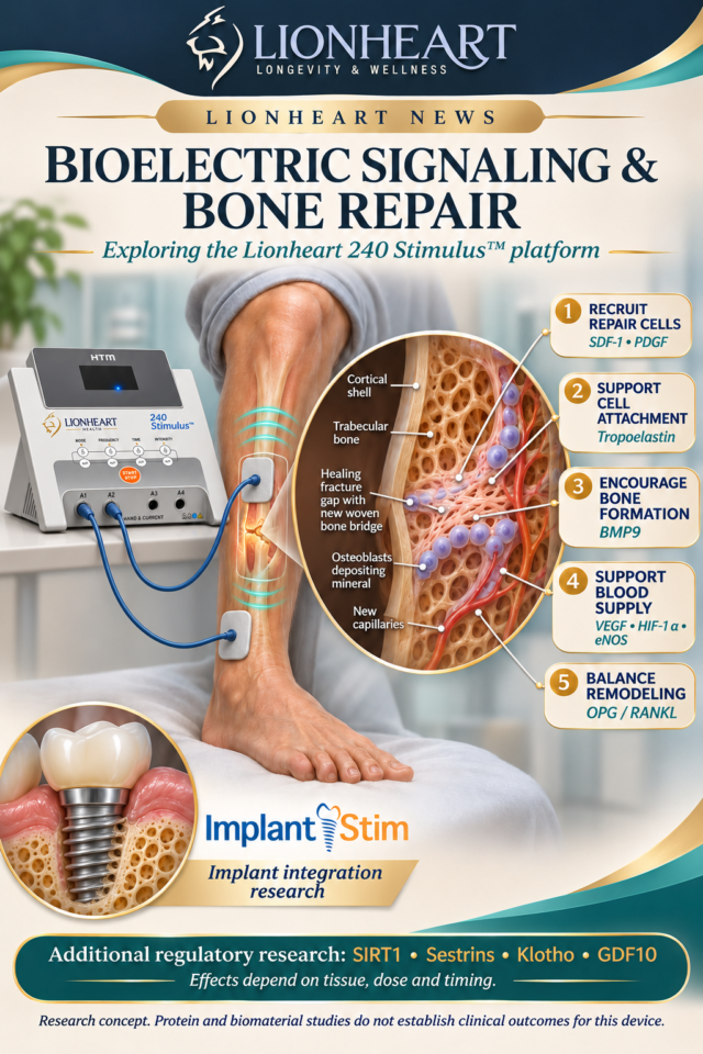
Tropoelastin is the soluble precursor of elastin, the protein that gives many tissues elasticity. It also acts as a biological signal. Laboratory research shows that tropoelastin can attract mesenchymal stem cells and stimulate their proliferation through interactions with cell-surface integrins αvβ3 and αvβ5. These findings suggest a potential role in recruiting and expanding cells needed for tissue repair.
Its relevance to bone is supported by experiments using tropoelastin-functionalized zirconium surfaces. Compared with bare zirconium, osteoblast-like cells showed approximately 30% greater attachment, up to 70% greater proliferation, twice the alkaline phosphatase production, and 30% greater mineral deposition. These are laboratory indicators of a more favorable bone-forming response. The experiments used osteosarcoma-derived osteoblast-like cells and coated materials; they were not clinical implant-healing trials.
For Lionheart’s ImplantStim™ research, this creates a biological rationale for investigating tropoelastin modulation around dental implants. It does not yet demonstrate that electrically induced tropoelastin produces the same results as a coating or reduces healing time in patients.
BMP9: Encouraging Cells to Build Bone
Bone morphogenetic protein 9, or BMP9, is a strong experimental driver of osteogenic differentiation—the process through which precursor cells become bone-forming osteoblasts.
In an osteoporotic rat fracture model, BMP9 delivered through an adenoviral approach increased callus formation and supported early mechanical stability. This provides preclinical evidence for BMP9 as a bone-repair target, although that delivery method differs from bioelectric stimulation.
Lionheart’s research concept is to investigate whether locally controlled BMP9 expression can help direct recruited repair cells toward bone formation. Demonstrating that effect requires measurements of protein expression, new bone formation, and mechanical strength.
OPG and RANKL: Balancing Bone Preservation and Remodeling
Bone repair needs both construction and controlled removal. Osteoblasts produce new bone, while osteoclasts resorb bone during remodeling.
RANKL promotes osteoclast formation and activity. Osteoprotegerin, or OPG, acts as a decoy receptor that limits RANKL signaling. Leonhardt’s published OPG patent documents describe bioelectric modulation of this pathway.
The potential application is stage dependent: excessive resorption may undermine bone preservation, but some osteoclast activity is necessary to remodel healing tissue. A useful protocol would therefore seek an appropriate balance rather than continuously suppressing bone turnover.
SDF-1 and PDGF: Recruiting and Expanding the Repair Team
Stromal cell-derived factor 1, also called SDF-1 or CXCL12, helps guide responsive cells toward sites where repair is needed. In a mouse study, locally delivered SDF-1 improved healing of critical-sized skull defects. Its effects differed between experimental models, emphasizing the importance of the surrounding tissue environment.
Platelet-derived growth factor, or PDGF, supports cell recruitment and proliferation. It also has human clinical evidence: a randomized periodontal study found that locally supplied recombinant PDGF-BB with a scaffold supported periodontal regeneration, including bone-related outcomes. Those findings concern a specific protein formulation and delivery system, rather than electrically induced PDGF expression.
Timing matters here, too. Experimental studies show that PDGF can inhibit BMP2-driven osteogenic differentiation under certain conditions. Expanding a population of repair cells and directing those cells to mineralize bone may require different signaling stages.
VEGF, HIF-1α, and eNOS: Supporting the Blood Supply
New bone needs oxygen, nutrients, and functioning blood vessels.
VEGF promotes blood-vessel formation. HIF-1α coordinates cellular responses to low oxygen and can activate angiogenic pathways. In rat fracture research, HIF-1α and VEGF increased within the developing callus, supporting their involvement in the healing response.
Endothelial nitric oxide synthase, or eNOS, contributes to nitric oxide signaling and vascular function. Research using endothelial-progenitor-cell conditioned media linked HIF-1α/eNOS activity with enhanced angiogenic properties; the conditioned media also improved vascularization and fracture healing in an animal model. This supports investigation of the pathway without proving that increasing eNOS alone produces the same benefit.
Together, these pathways offer a rationale for studying vascular support alongside bone-building signals.
Sirtuins, Sestrins, and Klotho: Regulating the Healing Environment
These proteins attract interest because of their roles in metabolism, cellular stress, and aging biology. Their skeletal effects require individual evaluation.
Sirtuins: SIRT1 has experimental evidence connecting it to bone-mass regulation and suppression of sclerostin, a protein that inhibits bone formation. This supports investigation of SIRT1-related signaling, although findings for one sirtuin cannot automatically be extended to the entire family.
Sestrins: Sestrin2 participates in cellular stress responses, but its bone effects are not uniformly protective. One study found that deleting Sestrin2 reduced RANKL-induced osteoclast formation, while osteoblast differentiation was unaffected. Increasing Sestrin2 therefore cannot presently be described as a proven way to strengthen or repair bone.
Klotho: Klotho is relevant to mineral metabolism and aging research, but more Klotho does not necessarily mean more bone formation. In cultured human bone-marrow mesenchymal stem cells, secreted Klotho reduced osteogenic differentiation. Any bone-repair application must account for protein form, concentration, tissue location, and treatment timing.
GDF10: A Potential Regulator, With Important Limits
Growth differentiation factor 10, or GDF10, should not be presented as an established bone-building signal.
Experimental research identifies GDF10 as an inhibitor of osteoblast differentiation and shows that it can reduce pathological mineral deposition in vascular tissues. Preventing calcification in an artery and encouraging mineralization within a fracture are different objectives. These findings make GDF10 an interesting regulatory research target, but they do not establish that increasing it at a bone injury improves repair.
Other Portfolio Proteins: Additional Opportunities and Unanswered Questions
Other proteins associated with the broader Leonhardt research portfolio may influence parts of the repair process:
-
Sonic hedgehog: Animal research supports a role in skeletal regeneration, including a requirement for full repair of large rib injuries in one mouse model. Its importance varies with injury size and setting.
-
FGF2: A specific low-molecular-weight FGF2 isoform accelerated fracture healing in transgenic mice, illustrating why the exact protein form matters.
-
GDF11, follistatin, and activins: These interacting pathways can influence muscle and bone differently. Published animal studies report conflicting GDF11 effects, and one study found that follistatin overexpression increased muscle mass while weakening bone. They require careful evaluation rather than a general claim of skeletal benefit.
Other portfolio targets—including IGF-1, HGF, EGF, TGF-β, collagen-related pathways, apelin, BDNF, NANOG, COL17A1, LIM proteins, and S100-family proteins—should be assessed individually before assigning them a bone-repair benefit. Evidence from skin, muscle, nerve, or aging research does not automatically establish improved fracture healing or implant integration. An exhaustive, current classification of every issued, pending, and in-process target would also require the company’s complete IP register.
What This Means for Lionheart’s Bone-Repair Research
The most promising concept is a coordinated sequence: recruit repair cells, support their survival and expansion, restore blood supply, encourage bone formation, and regulate remodeling.
Tropoelastin, BMP9, SDF-1, PDGF, OPG/RANKL, and vascular-support pathways provide meaningful biological reasons to investigate this approach. Klotho, sestrins, sirtuins, GDF10, and other regulatory proteins add opportunities—and important questions about dose, location, and timing.
For ImplantStim™ and related programs, the next scientific step is to connect measured bioelectric changes in protein expression with measured improvements in bone repair. Relevant outcomes include implant stability, imaging-confirmed bone formation, mechanical strength, time to safe loading, and adverse events.
Lionheart’s opportunity is to translate this complex biology into reproducible treatment protocols. The existing research supports that investigation; controlled studies must establish which signals, combinations, and sequences deliver meaningful benefits to patients.
Scientific References
These references support the biological mechanisms and limitations discussed in this article. They do not establish clinical effectiveness of Lionheart’s bioelectric protocols.
-
Yeo GC, Santos M, Kondyurin A, Liskova J, Weiss AS, Bilek MMM. Plasma-activated tropoelastin functionalization of zirconium for improved bone cell response. ACS Biomater Sci Eng. 2016;2(4):662–676. doi:10.1021/acsbiomaterials.
6b00049. -
Yeo GC, Weiss AS. Soluble matrix protein is a potent modulator of mesenchymal stem cell performance. Proc Natl Acad Sci U S A. 2019;116(6):2042–2051. doi:10.1073/pnas.1812951116.
-
Wang X, Huang J, Huang F, et al. Bone morphogenetic protein 9 stimulates callus formation in osteoporotic rats during fracture healing. Mol Med Rep. 2017;15(5):2537–2545. doi:10.3892/mmr.2017.6302.
-
Simonet WS, Lacey DL, Dunstan CR, et al. Osteoprotegerin: a novel secreted protein involved in the regulation of bone density. Cell. 1997;89(2):309–
319. doi:10.1016/S0092-8674(00) 80209-3. -
Jin Q, Giannobile WV. SDF-1 enhances wound healing of critical-sized calvarial defects beyond self-repair capacity. PLoS One. 2014;9(5). doi:10.1371/journal.pone.
0097035. -
Nevins M, Kao RT, McGuire MK, et al. Platelet-derived growth factor promotes periodontal regeneration in localized osseous defects: 36-month extension results from a randomized, controlled, double-masked clinical trial. J Periodontol. 2013;84(4):456–
464. doi:10.1902/jop.2012.120141. -
Wang X, Matthews BG, Yu J, et al. PDGF modulates BMP2-induced osteogenesis in periosteal progenitor cells. JBMR Plus. 2019;3(5). doi:10.1002/jbm4.10127.
-
Komatsu DE, Hadjiargyrou M. Activation of the transcription factor HIF-1 and its target genes, VEGF, HO-1, iNOS, during fracture repair. Bone. 2004;34(4):680–
688. PubMed PMID: 15050899. -
Kong L, Wang Y, Wang H, et al. Conditioned media from endothelial progenitor cells cultured in simulated microgravity promote angiogenesis and bone fracture healing. Stem Cell Res Ther. 2021;12:47. doi:10.1186/s13287-020-02074-y
. -
Cohen-Kfir E, Artsi H, Levin A, et al. Sirt1 is a regulator of bone mass and a repressor of Sost encoding for sclerostin, a bone formation inhibitor. Endocrinology.
2011;152(12):4514–4524. doi:10.1210/en.2011-1128. -
Oh SY, Kang N, Kang JY, et al. Sestrin2 regulates osteoclastogenesis via the p62-TRAF6 interaction. Front Cell Dev Biol. 2021;9:646803. doi:10.3389/fcell.2021.646803.
-
Zhang W, Xue D, Hu D, et al. Secreted klotho protein attenuates osteogenic differentiation of human bone marrow mesenchymal stem cells in vitro via inactivation of the FGFR1/ERK signaling pathway. Growth Factors.2015;33(5–6):356–365. doi:10.3109/08977194.2015.
1108313. -
Platko K, Gyulay G, Lebeau PF, et al. GDF10 is a negative regulator of vascular calcification. J Biol Chem.2024;300(11):107805. doi:10.1016/j.jbc.2024.107805.
-
Serowoky MA, Kuwahara ST, Liu S, Vakhshori V, Lieberman JR, Mariani FV. A murine model of large-scale bone regeneration reveals a selective requirement for Sonic Hedgehog. NPJ Regen Med. 2022;7:30. doi:10.1038/s41536-022-00225-8
. -
Hurley MM, Adams DJ, Wang L, et al. Accelerated fracture healing in transgenic mice overexpressing an anabolic isoform of fibroblast growth factor 2. J Cell Biochem. 2016;117(3):599–611. doi:10.1002/jcb.25308.
-
Liu W, Zhou L, Zhou C, et al. GDF11 decreases bone mass by stimulating osteoclastogenesis and inhibiting osteoblast differentiation. Nat Commun. 2016;7:12794. doi:10.1038/ncomms12794.
-
Suh J, Kim NK, Lee SH, et al. GDF11 promotes osteogenesis as opposed to MSTN, and follistatin, a MSTN/GDF11 inhibitor, increases muscle mass but weakens bone. Proc Natl Acad Sci U S A. 2020;117(9):4910–4920. doi:10.1073/pnas.1916034117.